


Files
Download Full Text (1.1 MB)
Date of Graduation
5-2027
Description
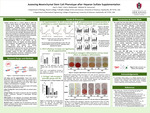
Multiple sclerosis is an autoimmune disease that damages the protective coating of nerves, often causing lasting neurological symptoms. Current treatments slow disease activity but rarely repair existing damage. In response to this, a novel treatment option has emerged: mesenchymal stem cell (MSC) therapy. MSCs are cells found in different adult tissues like bone marrow, and they have the potential to release repair-supporting signals and moderate inflammation. Nevertheless, results in a clinical setting have been inconsistent due to variations in cell potency. As a potential strategy to enhance MSC therapeutic potential, the addition of heparan sulfate, a naturally occurring signaling molecule, to cell cultures has been suggested. However, it is important that this molecule does not harm core MSC characteristics such as growth, multipotency, or cellular aging. Here, we investigate whether heparan sulfate alters MSC proliferation rate, colony forming ability, the rate at which these cells become senescent (or non-functional), ability to become other cell types, and the quantity of signaling molecules they secrete. Materials and Methods:Human bone marrow-derived MSCs from three different donors were purchased from a commercial vendor and expanded in cell culture media with or without the supplementation of heparan sulfate. These culture conditions were maintained for 3-4 passages. Subsequently, cell proliferation and colony forming ability were assessed using a Cell Counting Kit-8 (CCK) assay and a colony forming unit fibroblast (CFU-F) assay. Following that, the number of senescent cells was quantified through a Senescence-Associated β Galactosidase (SA-β-gal) staining assay. Next, cell differentiation into fat and bone cells was assessed by staining the oil droplets and calcium present in the cultures. Lastly, the amount of secreted cytokines and growth factors were measured by a multiplex assay using conditioned media. Results:The results from this study indicate that HS increases MSC proliferation most prominently in one donor, with minimal effects in the others, indicating a donor-dependent proliferative response. Moreover, the SA-β-gal assay showed that the percentage of senescent cells did not have a significant difference from MSCs treated with HS compared to those without treatment. Interestingly, HS seemed to increase the differentiation potential for both bone and fat cell types as indicated by the intensity of Alizarin Red and Oil-Red-O staining respectively. HS effects on colony-forming ability have been assessed. Consistent with these functional changes, HS-treated MSCs exhibited significantly higher inflammatory cytokines (IL-6, IL-8, MCP-1, IL-1α/β, TNFα) and growth factors (VEGF-A and PDGF) across all donors. Conclusion:This study demonstrates that HS increases MSC proliferative potential as well as their multipotency and secretion of different cytokines and growth factors without inducing cell senescence. These characteristics make HS a promising supplementation for MSCs since it does not negatively affect key stem cell characteristics.
Publication Date
2026
Document Type
Book
Degree Name
Bachelor of Science in Biomedical Engineering
Degree Level
Undergraduate
Department
Biomedical Engineering
Advisor/Mentor
Samsonraj, Rebekah
Disciplines
Biomedical Engineering and Bioengineering
Keywords
Engineering
Citation
Patel, I. (2026). Assessing Mesenchymal Stem Cell Phenotype after Heparan Sulfate Supplementation. 2026 Research Poster Competition. Retrieved from https://scholarworks.uark.edu/hnrcsturpc26/25