


Files
Download Full Text (1.9 MB)
Date of Graduation
12-2026
Description
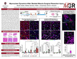
Displaced myonuclei are characteristic of skeletal muscle pathology and injury but are also found after exercise and with aging. These myonuclei are typically attributed to the incorporation of muscle stem cells (satellite cells).
To address whether displaced myonuclei in adult skeletal muscle originate strictly via outside sources such as satellite cells or can result from resident myonuclear migration, in regeneration conditions.
We used a muscle-fiber specific doxycycline-inducible resident myonuclear labeling mouse model (HSA-GFP, HSA = human skeletal actin promoter; GFP = green fluorescent protein) to fluorescently label and track resident myonuclei in adult mice (N=12, 10 months old). The lower third of the gastrocnemius was surgically removed, and the injury site of the remaining two thirds were analyzed at two timepoints (3 days and 7 days) post-surgery, as well as a non-injury control condition. 5-ethynyl-2’-deoxyuridine (EdU) was administered to track de novo DNA synthesis; displaced myonuclei and embryonic myosin positive (eMyHC+) fibers were quantified using immunohistochemistry, including fiber-type specific analysis. Data were analyzed using two-way ANOVAs with Tukey’s post-hoc tests.
The control and 3-day conditions did not significantly differ, as an average of 1.3% and 1.5% of fibers contained displaced nuclei, respectively. These displaced nuclei were predominantly GFP+/EdU- (resident) myonuclei, comprising 98% and 97% of the displaced myonuclear pool. By 7 days, there was a 6-fold increase (p < 0.01) in fibers containing a displaced nucleus compared to 3 days, at 8.89%, with 47% from GFP+/EdU- myonuclei and 53% from proliferated satellite cells (EdU+). When assessing fibers with multiple displaced myonuclei, the control and 3 day conditions on average had 0.09% and 0.17% of fibers containing multiple displaced nuclei, respectively, and were mostly GFP+/EdU-. By 7 days, there was an 18-fold increase in number of fibers containing multiple displaced nuclei compared to 3 days, with most of these coming from GFP-/EdU+ nuclei (74%). Fiber type-specific analysis revealed that the vast majority of displaced nuclei (~2/3) were in type IIB fibers, which is expected due to being the predominant fiber type in mouse gastrocnemius muscle. In the 7 day condition, an average of 5.8% of total fibers were positive for embryonic myosin expression.
These results challenge the dogma that displaced myonuclei are exclusively satellite cell derived during muscle injury/regeneration. Instead, myonuclear displacement in skeletal muscle following severe surgical trauma is due to both resident myonuclear migration and stem cell fusion. These data warrant further research into the role of resident myonuclear migration in skeletal muscle injury and reveal insights into possible therapies.
Publication Date
2026
Document Type
Book
Degree Name
Bachelor of Science in Exercise Science with Honors
Degree Level
Undergraduate
Department
Health, Human Performance and Recreation
Advisor/Mentor
Murach, Kevin
Committee Member
Rosa-Caldwell, Megan
Disciplines
Cells | Laboratory and Basic Science Research | Musculoskeletal System
Keywords
Research-Based
Citation
Goeke, M. A., Serrano, N., Koopmans, P. J., & Murach, K. A. (2026). Myonuclear Dynamics After Skeletal Muscle Surgical Resection Injury. 2026 Honors Symposium. Retrieved from https://scholarworks.uark.edu/coesym26/14
Included in
Cells Commons, Laboratory and Basic Science Research Commons, Musculoskeletal System Commons